
SPECT/CT Scans
A SPECT/CT scan pairs functional and anatomical imaging in one exam, helping your care team see both where activity is occurring in the body and exactly what anatomy it corresponds to. This combination makes SPECT/CT a valuable tool for diagnosis, staging, and treatment planning.
What is SPECT/CT Imaging?
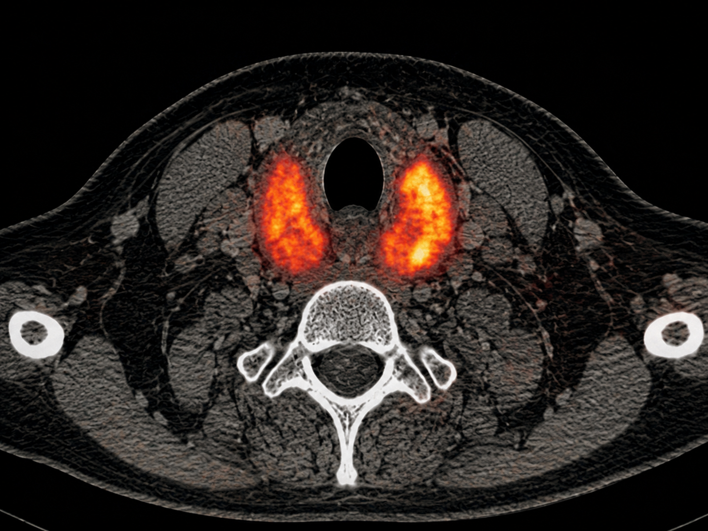
SPECT/CT stands for single-photon emission computed tomography combined with computed tomography. The SPECT portion shows how a radiotracer is distributed in the body to help identify areas of abnormal tissue activity, such as cancer or infection, while the CT portion helps localize that activity to specific anatomy.
This combination can help physicians evaluate both function and structure in a single imaging study. Depending on the clinical question, SPECT/CT may help localize disease activity, clarify findings seen on other imaging, evaluate treatment response, support surgical or therapy planning, or provide additional information when anatomical imaging alone is not enough.
Clinical Uses of SPECT/CT Imaging
SPECT/CT supports a broad range of evaluations across nuclear medicine. Depending on the radiotracer and clinical indication, it may be used to evaluate:
Bone metastasis and tumor evaluation
SPECT/CT can help localize abnormal radiotracer uptake to specific skeletal structures, supporting evaluation of suspected bone metastases, osseous lesions, and tumor-related bone involvement.
The CT component adds anatomical context that can help clarify whether uptake corresponds to degenerative change, trauma, tumor involvement, or another process.
Bone pain, trauma, and viability evaluation
SPECT/CT may support evaluation of focal bone pain, occult fracture, stress injury, postoperative change, and bone viability.
By combining functional uptake patterns with CT anatomy, SPECT/CT can help physicians better localize abnormalities and interpret findings that may be difficult to characterize on planar imaging alone.
Treatment response monitoring
In selected clinical settings, SPECT/CT may help physicians assess radiotracer distribution, disease activity, and changes over time after therapy.
This can be relevant for oncology follow-up, radiopharmaceutical therapy-related evaluation, and situations where functional imaging adds context beyond anatomical imaging alone.
Parathyroid adenoma localization
SPECT/CT can be used with appropriate radiotracers to support localization of parathyroid adenomas in patients being evaluated for hyperparathyroidism.
The fusion of nuclear medicine findings with CT anatomy may help localize abnormal parathyroid tissue and assist surgical planning when clinically appropriate.
Prosthetic joint evaluation
SPECT/CT may help evaluate painful or complicated prosthetic joints by localizing abnormal uptake around hardware and adjacent bone.
Depending on the clinical question and radiotracer protocol, it may support assessment of loosening, stress reaction, infection-related concerns, or postoperative changes.
Infection and inflammation imaging
SPECT/CT can help localize suspected infection or inflammatory activity when paired with the appropriate nuclear medicine study.
It may be useful in selected evaluations involving osteomyelitis, hardware-associated concerns, fever of unknown origin, inflammatory processes, or complex postoperative anatomy.
Cardiac perfusion imaging
In cardiac nuclear medicine, SPECT imaging can evaluate myocardial perfusion and support assessment of blood flow to the heart muscle.
When CT is incorporated, it can provide additional anatomical localization or attenuation correction depending on the equipment, protocol, and clinical indication.
Postoperative evaluations
SPECT/CT may be useful after surgery when anatomy has changed and conventional imaging is difficult to interpret.
By combining functional information with CT localization, SPECT/CT can help clarify whether radiotracer uptake corresponds to expected postoperative change, healing, complication, infection, or recurrent disease depending on the clinical context.
Thyroid, liver, and renal evaluation and treatment
SPECT/CT can support selected thyroid, liver, and renal nuclear medicine evaluations when anatomical localization improves interpretation.
Depending on the radiotracer and protocol, it may help localize functional findings, clarify equivocal planar imaging, or support treatment-related assessment.
Radiopharmaceutical therapy support
SPECT/CT can support selected radiopharmaceutical therapy workflows by helping evaluate radiotracer distribution, lesion localization, post-therapy uptake, and dosimetry-related questions.
This can be valuable when physicians need to understand where therapeutic activity is occurring and how it relates to patient anatomy.
Built for Referring Physicians Who Need Timely Answers
UniThera Medical Imaging is designed to support physicians who need responsive scheduling, reliable coordination, and clinically meaningful nuclear medicine imaging.
Our team helps referring offices move patients through the imaging process with fewer administrative barriers. Prior authorization is secured before the appointment whenever required, and appointments are typically available within 24 hours, with same-day scheduling when available.
For referring providers, SPECT/CT can be especially useful when the clinical question requires functional information, precise localization, or additional context beyond structural imaging alone.


What Patients Can Expect
A SPECT/CT appointment usually begins with a radiotracer given by injection, capsule, or another method depending on the exam. After the radiotracer is administered, there may be a waiting period while it distributes in the body. The timing varies by study and may range from a short interval to several hours.
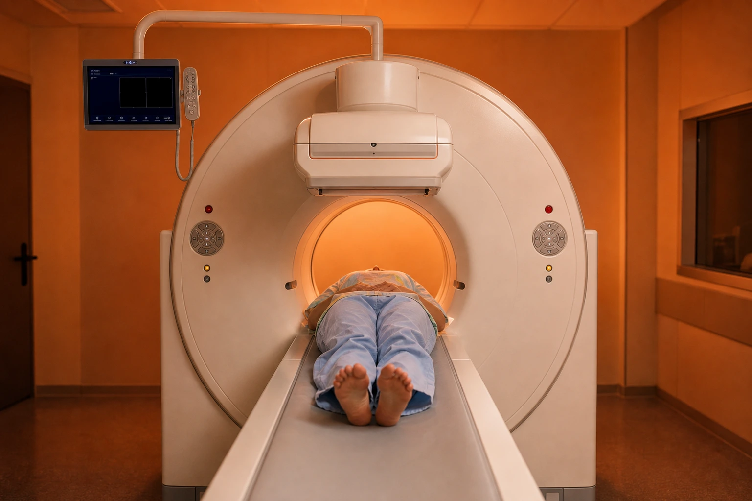
During the scan, the patient lies on a table while the imaging system captures nuclear medicine images and a CT scan for anatomical localization. The scan itself is noninvasive, and the technologist will provide instructions throughout the appointment.
Preparation instructions vary by exam. Patients will receive study-specific instructions before the appointment, including any guidance about food, medications, hydration, or arrival timing.
Standard of Care
Exceptional support before, during, and after imaging
UniThera Medical Imaging is designed to make advanced imaging easier to access, easier to coordinate, and easier to trust.
Scheduling
Appointments are typically available within 24 hours, with same-day scheduling when available.
Authorization
Our team secures prior authorization before every appointment.
Access
Direct, convenient parking is available steps from our front door.
Support
A clinical support team helps patients and referring physicians move imaging forward.
FAQ
Frequently Asked Questions
Answers to common questions about SPECT/CT imaging, scheduling, referrals, and what patients can expect.
What is SPECT/CT imaging?
SPECT/CT combines single-photon emission computed tomography (SPECT), which maps how radiotracers are distributed in the body, with computed tomography (CT), which creates detailed anatomical images.
How is SPECT/CT different from a regular CT scan?
A CT scan shows detailed anatomy. SPECT/CT adds functional nuclear medicine information, helping physicians understand where a radiotracer is active and how that activity relates to anatomy.
Is SPECT/CT only used for cancer?
No. SPECT/CT can support oncology care, but it is also used for bone, endocrine, cardiac, infection, inflammation, neurologic, and other nuclear medicine evaluations.
Do patients need a physician referral?
Most SPECT/CT studies require an order from a physician or qualified referring provider. Patients and referring offices can contact UniThera Medical Imaging for help with next steps.
How quickly can SPECT/CT imaging be scheduled?
Appointments are typically available within 24 hours, though insurance pre-authorization or other factors can extend that timeline. United Theranostics manages pre-authorization on our patient's behalf to minimize any potential delays.
Does the radiotracer stay in the body?
Radiotracers used for SPECT/CT are selected for the specific exam and generally leave the body over time, often through normal elimination pathways. Patients may be encouraged to hydrate after the exam, depending on the study.
What is the typical turnaround time for reporting/analysis?
We offer 24-48 hour turnaround on reports, so imaging doesn’t become a bottleneck in the treatment timeline. We also provide referring physicians with access to board-certified diagnostic radiology and nuclear medicine physicians for consultations before and after appointments.
SPECT/CT Availability
SPECT/CT imaging is available at most UniThera Medical Imaging locations. Availability may vary by site, protocol, and clinical indication.
View our locations to find the UniThera Medical Imaging site most relevant to your patient or appointment request.

Mailing Address:
United Theranostics
125 W South Street
Unit 3262
Indianapolis, IN 46206
Maryland
Crain Health Park
331 Oak Manor Drive
Suite 201
Glen Burnie, MD 21061
5454 Wisconsin Avenue
Suite 1600
Bethesda, MD 20815
New Jersey
Forrestal Medical Building
8 Forrestal Road South
Suite 201
Princeton, NJ 08540
New Mexico
4391 E Lohman Ave.
Suite A
Las Cruces, NM 88011